Day 1: Cell Surface Area
Preparation: Make a 2% agar solution by combining agar powder and about 2500 mL of boiling water. (This should be enough to fill a cake pan to a depth of 3 centimeters.) Stir the solution until all of the agar powder dissolves. As the agar solution cools, add 1 g of solid phenolphthalein (or 3 mL of liquid phenolphthalein) per liter of solution and stir well. The solution should be colorless. Pour into the cake pan and allow it to solidify.
Tell the class, “In this activity, we will use agar cubes to represent cells. The objectives for this activity are: to determine the surface area to volume ratio and relate this to cell size, in order to determine why cells divide, and to understand the problem cell growth causes and how cell division solves the cell growth problem.”
Have pairs of students put on goggles and gloves for the activity. Give each pair a 6 × 3 × 3-cm block of agar, a plastic knife, a metric ruler, and paper towels. Have them cut three cubes: 1 ×1× 1-cm, 2 × 2 × 2-cm, and 3 × 3 × 3-cm.
Ask students to place the cubes into the 250-mL beaker. Then, you should pour enough NaOH into the beaker to cover the cubes. Caution: NaOH is caustic and can burn the skin and eyes.
Leave the cubes in the NaOH solution for ten minutes, stirring occasionally. Explain that phenolphthalein is an indicator that will cause the agar cubes to turn pink when they are exposed to the basic NaOH solution.
While they are waiting, have students complete the data table (except for the “Distance the Solution Traveled into the Cube” column) and analysis questions on the Cell Surface Area worksheet (S-B-4-1_Cell Surface Area and KEY.docx).
After ten minutes, remove the agar cubes from the beaker with a spoon and dry them on a paper towel. Have students cut the cubes in half and measure the distance from the outer edge that has turned pink and record this on the worksheet.
Students will discover that the distance the solution diffused inward is the same for all the cubes. Ask them if the indicator represented a substance that the cell needed for survival, which “cell” got the needed substance throughout. They should answer that the smallest cell is pink throughout, so it is the most efficient. Have them relate the surface area-to-volume ratio to the results of the activity. (The smallest cube has the largest surface area-to-volume ratio. This shows that a large surface area-to-volume ratio promotes efficient distribution of substances and helps cells to survive.)
Explain the fact that the number of cells determines the size of an organism, and inform students, “Cells are limited in their growth for the sake of cell survival.” Explain that drawings and images under a microscope are simplified two-dimensional representations of cells, but actual cells are three-dimensional and much more complex. Inform students that, “Cells are so small because as the cell’s radius increases, the volume (amount of space/material within a cell) increases by an amount equal to the cube of the radius. What that means is that if the radius of a cell doubles, the volume increases by a factor of 8; if the radius of a cell triples, the volume increases by a factor of 27, and so on.”
Write on the board:
23 = 8
33 = 27
Ask, “If the radius of a cell was to increase by a factor of 5, by what factor would its volume increase?” Give students about 1 minute to come up with the answer within their groups. Have each group share its answer. (53 = 125)
Explain that cells must absorb nutrients in order to survive, and the amount of nutrients needed depends on the volume. If the volume increases by a factor of 8, the amount of nutrients the cell needs to absorb increases by a factor of 8, and so on. Because of this, there is a size limit for cells; they reach a point at which they are unable to grow any larger. The cell cycle is essential so that cells can grow and divide to be efficient.
To assess students’ understanding of the relationship between surface area to volume and cell size, have students write a paragraph in conclusion to the activity by answering the question: “Which cell would have a better chance for survival, one that is 1 × 1 × 1-cm or one that is 0.1 × 0.1 × 0.1-cm? Why?”
Day 2: The Cell Cycle
Preparation: Make copies of Cell Cycle Stages (S-B-4-1_Cell Cycle Stages.doc). Cut out the stages and tape or glue them to index cards. Color-code the index cards with dots using markers but do not write the names of the stages on the cards (see key below). Make a set for each group of five students. Index Card Key:
- Red—G0
- Orange—G1
- Yellow—S phase
- Green—G2
- Blue—M phase
Ask students to name some events that happen in cycles. Write their answers on the board. Examples include the cycle of the seasons, analog clocks (the hands move through two separate 12-hour cycles per day), and the wash cycle on a washing machine. Say to the class, “Now that we have some examples of events that occur in cycles, think about what a cycle is and write a definition of the word cycle in your notebook.” Allow 2–3 minutes and afterward, write the following definition of cycle on the board and have students copy it in their notes, comparing it to their own definition: “A cycle is a sequence of events, stages, or phases that repeats with some regularity in the same order.” Explain to students that the cell cycle is related to the growth and maximum size of cells.
Tell the class, “Now we will examine the cell cycle.” Explain, “The cell cycle is an ordered set of events that cells need for growth as well as division into daughter cells. Eukaryotic cells must replicate their genomes (DNA) and separate the replicated genome before they can divide.”
Write the heading CELL CYCLE on the board. Under the heading, write the following notes and instruct students to copy them. Read each note before you present it to students:
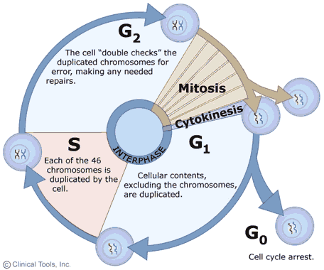
1. Three main stages: interphase, then nuclear division (mitosis) and cytokinesis.
2. Interphase is the longest stage, generally lasting 12 to 24 hours in mammal cells.
3. During interphase, the cell is constantly synthesizing RNA.
4. During interphase, the cell produces protein and grows.
5. Interphase can be divided into four steps: Gap 0 (G0), Gap 1 (G1), S (synthesis) phase, Gap 2 (G2).
6. Interphase ends when the M (mitotic) phase begins.
7. The mitotic phase includes mitosis, which includes prophase, metaphase, anaphase, and telophase.
8. Mitosis is the stage during which cells segregate duplicated chromosomes.
9. Cytokinesis occurs after mitosis.
10. During cytokinesis, the cytoplasm divides and one cell becomes two individual cells as they break apart. In animal cells, the two developing nuclei are separated as the cell constricts and forms two daughter cells. In plant cells, a new cell wall develops between the daughter cells.

Source: www.cbu.edu/~seisen/CellCycle_files/image001.gif
Explain, “Cells appear to be resting during interphase, but many activities occur during this period that make mitosis possible.”
Introduce the cell cycle sequencing activity. Hand out a set of 5 color-coded cards to each group and have them distribute the cards to the members of the group. Working as a group, students are to decide the proper order for the cards and arrange them in order. Give students about 5-7 minutes to read the cards, discuss what each person has on his/her card, and guess the correct order. After the time limit has expired, have each student group give the sequence that they think the cards should be in. After all groups have shared, tell students to look at the colored dot on each card and give them the correct order:
- Red—G0
- Orange—G1
- Yellow—S phase
- Green—G2
- Blue—M phase
Inform them that not all cells go through the G0 stage. For cells in which G0 occurs, it happens after G1 begins but before it ends.
Emphasize that not all cells undergo cell division, and that this cell cycle only applies to cells that divide.
End the lesson by having students complete the Cell Cycle Exit Ticket (S-B-4-1_Cell Cycle Exit Ticket and KEY.doc) containing the following directions:
- List the stages of interphase in order. (G1, S, G2, and in some cells, G0)
- What two phases make up the M phase of the cell cycle? (mitosis and cytokinesis)
- During which stage of interphase is DNA replicated? (S)
- Students who might need an opportunity for additional learning can view the animation at the site www.cellsalive.com/cell_cycle.htm and copy the circular diagram of the cell cycle stages.
- Students who might need an opportunity for additional learning can view the animations at the following Web sites and take the quizzes at the bottom.
Extension:
o http://highered.mcgraw-hill.com/sites/0072495855/student_view0/chapter2/animation__control_of_the_cell_cycle.html
